Analysis of periodontal disease in the archaeological population of Dąbrówki (Poland) (16th–17th centuries)
Institute of Biological Sciences, Cardinal Stefan Wyszynski University in Warsaw, Poland
 https://orcid.org/0000-0002-8163-961X
Department of Environmental Hazard Prevention and Allergology, Medical University of Warsaw, Poland
https://orcid.org/0000-0002-0605-665X
Institute of Biological Sciences, Cardinal Stefan Wyszynski University in Warsaw, Poland
Abstract
Introduction
Periodontitis is a disease affecting a significant proportion of both modern and historical populations, and its development is largely associated with poor oral hygiene.
Study aim
The aim of this study was to assess periodontal status of the historical population of Dąbrówki (Poland), dated to the 16th–17th centuries.
Material and Methods
The study sample consisted of dental material from 24 individuals (12 females and 12 males), divided into three age groups (17–25, 26–35, and >36 years). The analysis was based on measurement of the distance between the cementoenamel junction and the surface of the alveolar bone, assessment of changes in the mesial and distal interdental septa, evaluation of architectural changes in the alveolar bone, and assessment of the possible presence of molar furcation.
Results
Periodontitis was diagnosed in 75% (18/24) of the examined individuals, and pathological changes were identified in 28% (146/520) of the analysed teeth. The occurrence of the disease was related to sex, with periodontitis being statistically more frequent in males (46%) than in females (12%). An increase in disease severity with age was also observed.
Conclusions
The results indicate that the population of Dąbrówki exhibited a lower frequency of periodontitis than other Polish populations from a similar historical period. One possible explanation is the rural character of the community, where dietary patterns may have differed from those of urban populations. Research on the Dąbrówki population is ongoing, and further analyses, including isotopic studies, are expected to allow a more comprehensive interpretation of the findings.
Keywords: periodontitis, dental material, ancient Poland, oral hygiene, diet
Introduction
Periodontal disease is a persistent inflammation of the tissues that make up the periodontium, which may include the gums, alveolar bone, root cementum, and periodontal ligament (e.g., AAP, 2003; Armitage, 2004; Ogden, 2008). Periodontal diseases are divided into gingivitis and periodontitis. Gingivitis results from plaque accumulation, which leads to inflammation of the soft tissues around the tooth; this condition is reversible (e.g., Trombelli, 2025). Periodontitis, on the other hand, is an advanced form of the disease in which permanent damage to the periodontal structures occurs. Periodontal disease affects a significant proportion of the population worldwide. It is estimated that more than 42% of adults aged >30 years suffer from some form of gum disease, and nearly 8% have severe periodontal disease (e.g., Nascimento et al., 2024). In many individuals, the disease leads to tooth loss due to reduced stability and damage to the alveolar bone (Armitage, 1999; Highfield, 2009).
The main cause of periodontal disease is poor oral hygiene. As the disease progresses, anaerobic bacteria such as Porphyromonas gingivalis and Prevotella intermedia gradually replace the natural saprophytic microflora of a healthy mouth. Substances produced by these bacteria trigger an inflammatory response in the body, leading to damage to the tissues surrounding the tooth (Czerniuk et al., 2016; Li et al., 2025; Mehrinia & Van Dyke, 2026; Nazir, 2017; Pihlstrom et al., 2005). Some authors attribute periodontal disease to dietary habits, such as vitamin or micronutrient deficiencies (e.g. Garcia et al., 2001). Studies by Šlaus et al. (1997) suggest that greater access to highly cariogenic foods contributes to periodontal disease. However, the role of diet in periodontal disease remains under discussion (e.g. Balice et al., 2025). Other causes have also been identified; metabolic disorders, such as diabetes or insulin resistance, can exacerbate inflammatory processes in periodontal tissues (e.g. Saito & Shimazaki, 2007). In addition, excessive and chronic psychological stress (Aleksejūnienè et al., 2002; Genco et al., 1999) and the use of stimulants, such as smoking (Nociti et al., 2015), are recognised as contributing factors to periodontitis.
Studies of periodontitis are rarely conducted on historical populations because they require well-preserved teeth and dental arches (e.g., Karkus, 2018; Vodanović et al., 2012). Because of the disappearance of soft tissues after death, analyses of periodontal disease are usually limited to assessing the presence or absence of periodontitis in individuals (e.g., DeWitte & Bekvalac 2011; Kwiatkowska & Nowakowski, 2011; Oztunc et al., 2006). Other aspects, such as disease severity, are therefore often omitted. Nevertheless, studies of historical populations are valuable because they provide insight into the hygiene and dietary practices of past communities.
The most recent excavations, conducted between 2022 and 2024 at the Dąbrówki site in Poland, provide an opportunity to analyse this population with respect to periodontal disease and to use the resulting data to reconstruct local dietary and hygiene habits. The key research questions address the following issues: (i) What was the incidence rate of periodontal disease in the Dąbrówki population compared with other groups in Poland? (ii) Are there differences in incidence between the sexes? (iii) Can stages of disease progression be identified in the studied population?
Material and methods
The material used for this research derives from a cemetery in Dąbrówki, Podlaskie Province, Poland (Fig. 1). The cemetery was probably established for the burial of the local rural population. Because of the rare mention of rural burials in written sources, this cemetery – like most sites of this type – was discovered accidentally during construction work carried out in the vicinity of the village. Nevertheless, a small number of historical sources refer to the settlement as a place associated with Queen Bona and Sigismund Augustus (Wawrzeniuk, 2021). In several graves, artefacts were recovered (e.g. a silver coin bearing the image of Sigismund III Vasa dated to 1593), allowing for a preliminary dating of the cemetery to the 16th and 17th centuries. Radiocarbon (14C) dating indicates that the burials date to the period 1505–1615 AD (Wawrzeniuk et al., 2023).
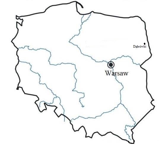
During the archaeological investigations, 62 graves were uncovered. However, because of the quality of preservation – particularly in the dental arch region – and the possibility of reliable sex and age determination, only 24 adult individuals (12 males and 12 females) were ultimately included in the analysis. In total, 520 permanent teeth were examined (226 anterior and 294 posterior). Only fully erupted teeth with antagonists in the opposing dental arch were included in the analysis (Table 1).
| Maxilla | ||||||||||||||||
|---|---|---|---|---|---|---|---|---|---|---|---|---|---|---|---|---|
| FDI | 11 | 12 | 13 | 14 | 15 | 16 | 17 | 18 | 21 | 22 | 23 | 24 | 25 | 26 | 27 | 28 |
| No. | 14 | 15 | 19 | 17 | 18 | 16 | 10 | 8 | 13 | 15 | 19 | 17 | 14 | 11 | 12 | 9 |
| Mandible | ||||||||||||||||
| FDI | 31 | 32 | 33 | 34 | 35 | 36 | 37 | 38 | 41 | 42 | 43 | 44 | 45 | 46 | 47 | 48 |
| No. | 22 | 22 | 23 | 21 | 19 | 14 | 18 | 8 | 20 | 22 | 22 | 21 | 18 | 13 | 17 | 13 |
FDI World Dental Federation (Fédération Dentaire Internationale, FDI)
The sex of individuals was determined on the basis of pelvic bone morphology as well as cranial traits, in accordance with the scoring system for sexually dimorphic cranial features proposed by Buikstra and Ubelaker (1994). Age at death was estimated using changes in the topography of the auricular surface of the ilium and morphological characteristics of the pubic symphysis.
Given the correlation between the incidence of periodontal disease and age, the sample was divided into three age categories: 17–25, 26–35, and >36 years. A similar age division has been used in previous research, allowing for comparability with other studies (Tomczyk et al., 2018).
The American Academy of Periodontics (AAP) and European Federation of Periodontology (EFP) indicate that the severity of periodontitis should be determined based on clinical attachment loss, pocket depth, bleeding on probing, and bone loss. According to these criteria, a patient has periodontitis when these symptoms are diagnosed (e.g., Chmielewski et al., 2025). However, the criteria cannot be used in the diagnosis of individuals from historical populations. Therefore, other methods of identifying periodontal disease and its severity are proposed. The following criteria were used for a reliable assessment of the presence of periodontal disease:
- Measurement of the distance between the cementoenamel junction (CEJ) and the surface of the alveolar crest (AC). Measurements were taken using callipers and recorded in millimetres (accuracy: 0.01 mm). A CEJ–AC distance of 3 mm is generally regarded as normal, whereas a distance of >3 mm is considered indicative of a lesion (e.g. Shaju, 2011).
- Assessment of changes in the mesial and distal interdental septa (interproximal bone) of each tooth. Observations were made at 10× magnification under standardised lighting conditions using an endodontic microscope (Global Surgical Corporation, USA). Findings were recorded as the presence (1) or absence (0) of the interdental septum.
- Evaluation of changes in the texture and architecture of the alveolar bone. For this criterion, the method proposed by Kerr (1988) was applied, with modifications introduced by our research team (Tomczyk et al., 2017). This classification allows the severity of periodontitis to be identified and divided into mild (early), moderate, and severe (advanced) stages.
- In cases where bi- or trifurcation of the root was exposed, the presence (1) or absence (0) of furcation involvement was assessed using a periodontal probe.
Periodontal disease was diagnosed only when all three criteria (i–iii) were met for anterior and posterior teeth. The fourth criterion (iv) was additionally introduced in accordance with the newest American Academy of Periodontics (AAP) and European Federation of Periodontology (EFP) recommendations for diagnosing moderate and advanced stages of periodontal disease (e.g., Chmielewski et al., 2025).
The presence of dental calculus was treated as an additional, but not essential, criterion for evaluation (e.g. Lieverse, 1999; Vodanović et al., 2012), as calculus on archaeological material may be easily damaged or lost during excavation, handling, or transport to the laboratory.
Statistical analyses were conducted using the R Project for Statistical Computing (version 4.3.1). Two-sample and three-sample tests of proportions were applied to identify significant differences between age groups and between females and males. Tables 2 and 3 report the chi-square test statistics and corresponding p-values. Differences with p ≤ 0.05 were considered statistically significant.
Results
The study included a total of 24 individuals (12 males and 12 females) with well-preserved dental and skeletal material. Pathological changes were identified in 18 individuals (9 males and 9 females), accounting for 75% of the analysed sample. In total, 520 teeth were examined, including 226 anterior and 294 posterior teeth. Evidence of periodontal disease was observed in 146 teeth (28%), of which 55 (24%) were anterior teeth and 91 (31%) were posterior teeth.
The results indicate variation in disease incidence across age groups. In the 17–25-year age group, periodontal disease was least frequent, affecting only 9% of the teeth examined. The highest prevalence was recorded in the 26–35-year age group, in which periodontal disease was identified in 44% of teeth; in this group, the disease occurred exclusively in males. Among individuals aged over 36 years, the prevalence of lesions was 39% and was significantly higher in males (45%) than in females (16%). Regardless of age group, periodontal disease was statistically more common in males (Table 2).
Because disease severity is likely to be related to age, this relationship was examined in the next stage of analysis (Fig. 2). Across all age groups and in both sexes, moderate periodontal disease was the most frequently observed stage. However, a marked increase in advanced disease was noted between the youngest (17–25 years) and middle (26–35 years) age groups (5% versus 30%) (Table 3).
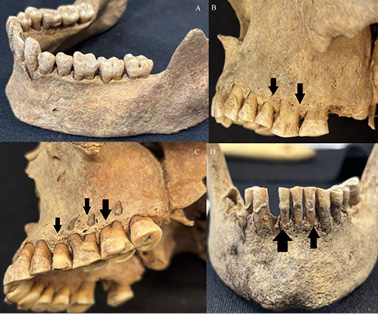
Differences in susceptibility to periodontal disease were also observed between the sexes. Among males aged 26–35 years, the moderate stage of periodontal disease predominated (40% versus 33%), whereas among females in the same age group, the early stage was most common (47% versus 27%) (Table 3).
| Age of individual | Total | Male | Female | p | χ2 |
|---|---|---|---|---|---|
| 17–25 | 19/212 (9%) | 7/73 (10%) | 12/139 (9%) | 1 | 1.7 |
| 26–35 | 67/154 (44%) | 52/52 (100%) | 15/102 (15%) | <0.0001 | 101.9 |
| >36 | 60/154 (39%) | 55/123 (45%) | 5/31 (16%) | 0.014 | 8.508 |
| p | 0.0001 | 0.0001 | 0.3032 | ||
| χ2 | 65.55 | 100.1 | 2.39 | ||
| Total | 146/520 (28%) | 114/248 (46%) | 32/272 (12%) | <0.0001 | 75.1 |
| Age of individual | Total | Male | Female | ||||||
|---|---|---|---|---|---|---|---|---|---|
| Early | Moderate | Advanced | Early | Moderate | Advanced | Early | Moderate | Advanced | |
| 17–25 | 2/19 11% |
16/19 84% |
1/19 5% |
– | 7/7 100% |
– | 2/12 17% |
9/12 75% |
1/12 8% |
| 26–35 | 21/67 31% |
26/67 39% |
20/67 30% |
14/52 27% |
21/52 40% |
17/52 33% |
7/15 47% |
5/15 33% |
3/15 20% |
| >36 | 22/60 37% |
33/60 55% |
5/60 8% |
22/55 40% |
31/55 56% |
2/55 4% |
– | 2/5 40% |
3/5 60% |
| p | 0.098 | 0.002 | 0.002 | 0.220 | 0.008 | 0.0002 | 0.218 | 0.088 | 0.062 |
| χ2 | 4.64 | 12.75 | 12.36 | 1.50 | 9.68 | 13.52 | 1.51 | 4.86 | 5.57 |
In the diagnosis of periodontitis, particular attention is often paid to measurements of the distance between the CEJ and the AC. The present study shows that this distance increases with disease progression. The lowest values (mean: 2.5 mm) were recorded in the healthy group, while mean measurements increased progressively in subsequent stages of the disease. An accompanying increase in standard deviation was also observed, indicating greater variability in CEJ–AC distances in more advanced stages of periodontitis (Table 4).
| Stage of periodontitis | min | max | mean | median | SD |
|---|---|---|---|---|---|
| Healthy | 1.0 | 7.1 | 2.5 | 2.2 | 1.06 |
| Early | 3.2 | 7.1 | 4.5 | 4.2 | 0.93 |
| Moderate | 4.0 | 9.6 | 6.3 | 6.1 | 1.13 |
| Advanced | 5.0 | 12.0 | 8.0 | 7.8 | 1.51 |
Discussion
The population studied from the Dąbrówki site shows a frequency of periodontal disease, in terms of the number of affected individuals, similar to that reported for other populations from Poland dated to a comparable historical period (16th–17th centuries) (Table 5). It may therefore be assumed that the environmental factors most commonly associated with periodontal disease, such as diet quality and oral hygiene, were broadly similar in the populations considered. However, it should be noted that apparent similarities in disease frequency may also result from differences in diagnostic methodology. In many earlier studies, authors relied on only one or two diagnostic criteria, such as visual assessment of alveolar bone resorption, the presence of dental calculus (Gleń, 1976), or CEJ–AC measurements alone (Kozubkiewicz & Trachtenberg, 1960). As a result, their estimates may be overstated. Only studies of the populations from Brześć Kujawski (Karkus, 2018) and Radom (Tomczyk et al., 2018) applied methods comparable to those used in the present research. For this reason, comparisons were limited to these two Polish populations.
Analysis of data from the chronologically similar populations of Brześć Kujawski and Radom (16th–17th centuries) suggests that the proportion of teeth affected by periodontal disease in the Dąbrówki population was relatively low (28%).
| Period | Site | Individuals | References |
|---|---|---|---|
| 15th-18th | Brześć Kujawski | 48/61 (79%) | Kozubiewicz & Trachtemberg 1960 |
| 14th-17th | Brześć Kujawski | 37/48 (77%) | Karkus 2018 |
| 15th-18th | Cracow | 68/108 (63%) | Gleń 1976 |
| 16th-18th | Wrocław | 18/30 (60%) | Kwiatkowska & Nowakowski 2011 |
| 14th-17th | Radom | 57/80 (71%) | Tomczyk et al. 2018 |
| 18th-19th | Radom | 93/126 (74%) | Tomczyk et al. 2018 |
| 16th-17th | Dąbrówki | 18/24 (75%) | presented publication |
In Brześć Kujawski, pathological changes were identified in 41% of examined teeth (Karkus, 2018), while in the population of Radom, dated to the late Middle Ages and early modern period (14th–17th centuries), periodontal disease was diagnosed in 44% of teeth (Tomczyk et al., 2018). The observed differences between the Dąbrówki population and the groups from Brześć Kujawski and Radom may be related to differences in socio-economic status. The population examined in the present study derives from a rural context, whereas the populations from Brześć Kujawski and Radom were associated with urban centres. Comparative studies indicate that during the late Middle Ages and early modern period, urban populations generally exhibited poorer oral health, which has been linked, among other factors, to diets richer in highly processed carbohydrates (e.g., Šlaus et al., 1997; Tomczyk et al., 2018). These factors may therefore help explain the higher incidence of periodontal disease observed in the urban populations. At the current stage of research, no independent data are available to confirm the quality or composition of the diet in the Dąbrówki population, as analyses of the material are still ongoing. By contrast, detailed studies conducted at the Radom site, including isotopic, archaeozoological, and archaeobotanical analyses, confirm the proposed pattern, namely that urban populations had poorer diets and a higher proportion of individuals affected by diseases of the masticatory system (Tomczyk et al., 2018; 2020).
Of course, it should be remembered that periodontitis is a multifactorial disease (e.g., Muro et al., 2026). Its development may be influenced by behavioural factors (lifestyle), nutritional factors, or metabolic disorders (such as diabetes or insulin resistance). However, the interpretation of results based on bone (archaeological) material is always limited to those elements that can be verified by scientific evidence. Therefore, when looking for the causes of periodontal disease in historical populations, the most common factors pointed to are diet quality and the socioeconomic status of the population studied.
The results obtained for the Dąbrówki population also reveal a clear contrast between males and females in the prevalence of periodontitis. Contemporary studies consistently show that periodontal disease is more common in men than in women (e.g., Ioannidou, 2017; Stănescu et al., 2020), and similar patterns have been documented in historical populations (e.g., Karkus, 2018; Novak, 2015; Raitapuro-Murray et al., 2014). Sex is considered a potential risk factor, as the composition of the oral microbiome differs between women and men, and sex hormone levels – particularly male androgens – may influence the development and progression of periodontal disease (e.g., Del Pinto et al., 2024; Mascarenhas et al., 2003). Clinical studies further indicate that oestrogen slows the production of inflammatory cytokines, such as interleukin-1β and tumour necrosis factor-α, which are responsible for bone resorption during periodontal inflammation. As a result, women tend to produce fewer cytokines in response to infection. Progesterone also plays an important role in the formation of periodontal ligaments and the maintenance of bone mass (e.g., Ahmed et al., 2010). These biological mechanisms may help explain the lower incidence of periodontal disease observed among women.
Dietary factors may also have contributed to sex-related differences in disease prevalence. Research by Reitsema et al. (2010) indicates that males in historical populations more frequently consumed meat products. Diets rich in protein are thought to promote dental calculus accumulation (Hillson, 1979), as increased protein intake leads to elevated blood urea levels. Oral bacteria metabolise urea into ammonia, resulting in increased oral alkalinity (Lieverse, 1999) and, consequently, greater calculus formation. These processes may provide an additional explanation for the significantly higher prevalence of periodontitis observed among males from Dąbrówki.
When discussing periodontal disease, the relationship between age and disease severity must also be considered. Periodontitis develops gradually, with its effects accumulating over time (e.g., Benatti et al., 2009; Flemmig, 1999). In the Dąbrówki population, a clear progression was observed between successive age groups (9% versus 44%). In the oldest age group, however, the prevalence of disease decreased to 39%. This phenomenon has been described in previous studies and is likely related to tooth loss in individuals affected by the most advanced stages of periodontitis (DeWitte & Bekvalac, 2011). With increasing age, changes in immune function may further exacerbate chronic inflammation and contribute to declining oral health (Lopez et al., 2017; Preshaw et al., 2012). Notably, disease progression appeared to occur more rapidly in males. In the 26–35-year age group, 40% of males were diagnosed with moderate periodontitis and 32% with advanced disease, whereas among females of the same age, 33% exhibited moderate disease and 20% advanced disease.
In odontological research, the use of multiple diagnostic criteria is essential. For many years, CEJ–AC measurement was the primary method for assessing periodontitis in skeletal material (e.g., DeWitte, 2012; Kozubkiewicz & Trachtenberg, 1960; Raitapuro-Murray et al., 2014). It is now recognised, however, that this criterion alone is insufficiently reliable and may lead to misdiagnosis (e.g. Tomczyk et al., 2017). Recent studies (e.g., Muro et al., 2026) suggest that macromorphological bone defects provide a more robust indicator of periodontal disease. For example, research conducted on a forensic collection from Yucatán demonstrated that bone changes were present in 94.8% of individuals, whereas CEJ values exceeded diagnostic thresholds in only 55.8% (Muro et al., 2026). Reliance solely on CEJ measurements would therefore result in substantial underestimation of disease prevalence. Although CEJ values correlate with disease severity, several authors emphasise that this measure should be used as a supportive tool rather than as a primary diagnostic criterion (Muro et al., 2026; Tomczyk et al., 2017). In the Dąbrówki population, the progressive increase in mean CEJ values across disease stages (Table 4) indicates that this measure retains comparative value, but it should be applied in conjunction with the assessment of bone defects to ensure diagnostic reliability and a more comprehensive interpretation of periodontal changes.
Limitations
The main limitation of the present study is the relatively small number of individuals included in the analysis. However, as noted above, research on periodontal disease requires not only well-preserved alveolar bone but also the presence of opposing teeth, which substantially restricts sample size in archaeological contexts.
Conclusions
Periodontal research is rarely represented in anthropological literature, largely because of the condition of preserved skeletal material. Additional challenges arise from the difficulty of accurately diagnosing periodontal disease in archaeological populations. Consequently, each contribution in this area is valuable because it can provide important insights into the dietary practices and hygiene habits of historical communities.
Although examinations of periodontal disease cannot always be conducted because of the preservation state of archaeological material, they can yield informative and meaningful results. The study of the Dąbrówki population indicates a lower frequency of periodontitis compared with other Polish populations from a similar historical period, a pattern that may be linked to the rural character of the community. Nevertheless, further research is required to confirm this interpretation. As discussed, the principal limitation of the study lies in the limited number of individuals analysed; however, this constraint is inherent to periodontal research, which depends on the preservation of both alveolar bone and opposing teeth.
References
(AAP) American Academy of Periodontology. (2003). Diagnosis of periodontal diseases. Journal of Periodontology, 74, 1237–1247. https://doi.org/10.1902/jop.2003.74.8.1237
Ahmed, S. A., Karpuzoglu, E., & Khan, D. (2010). Effects of sex steroids on innate and adaptive immunity. In: S. L. Klein & C. Roberts (Eds.), Sex Hormones and Immunity to Infection (pp.19–51). Springer: Berlin.
Aleksejūnienè, J., Holst, D., Eriksen, H. M., & Gjermo P. (2002). Psychosocial stress, lifestyle and periodontal health. A hypothesized structural equation model. Journal of Clinical Periodontology, 29, 326–335. https://doi.org/10.1034/j.1600-051x.2002.290408.x
Armitage, G. C. (1999). Development of a classification system for periodontal disease and conditions. Annals of Periodontology, 4, 1–6. https://doi.org/10.1902/annals.1999.4.1.1
Armitage, G. C. (2004). Periodontal diagnoses and classification of periodontal disease. Periodontology 2000, 34, 9–21. https://doi.org/10.1046/j.0906-6713.2002.003421.x
Balice, G., Paolantonio, M., Murmura, G., Serroni, M., Di Gregorio, S., & Femminella, B. (2025). The Influence of diet and physical activity on periodontal health: a narrative review. Dentistry Journal https://doi.org/10.3390/dj13050200
Benatti, B. B., Silvério, K. G., Casati, M. Z., Sallum, E. A., & Nociti, F. H. (2009). Inflammatory and bone-related genes are modulated by aging in human periodontal ligament cells. Cytokine, 46, 176–181. https://doi.org/10.1016/j.cyto.2009.01.002
Buikstra, J. E., & Ubelaker, D. H. (1994). Standards for data collection from human skeletal remains. Arkansas Archeological Survey Research Series, No.44. Arkansas: Fayetteville.
Chmielewski, M., Pilloni, A., Cuozzo, A., D’Albis, G., D’Elia, G., Papa, P., Marini, L. (2025) The 2018 Classification of Periodontitis: Challenges from Clinical Perspective. Dentistry Journal, 13. https://doi.org/10.3390/dj13080361
Czerniuk, M. R., Bartoszewicz, Z., Filipiak, K. J., Dudzik-Niewiadomska, I., Pilecki, T., & Górska, R. (2017). Plasmatic NT-proBNP concentrations in patients with coexistent periodontal disease and congestive heart failure: pilot studies. Kardiologia Polska, 75, 135–142. https://doi.org/10.5603/KP.a2016.0148
Del Pinto, R., Ferri, C., Giannoni, M., Cominelli, F., Pizarro, T. T., & Pietropaoli, D. (2024). Meta-analysis of oral microbiome reveals sex-based diversity in biofilms during periodontitis. JCI Insight, 10, 9(17), e171311. https://doi.org/10.1172/jci.insight.171311
DeWitte, S. N., & Bekvalac, J. (2011). The Association between periodontal disease and periosteal lesions in the St. Mary Grace Cemetery, London, England A.D.1350-1538. American Journal of Physical Anthropology, 146, 609–618. https://doi.org/10.1002/ajpa.21622
DeWitte, S. N. (2012). Sex differences in periodontal disease in catastrophic and attritional assemblages from medieval London. American Journal of Physical Anthropology, 149, 405–416. https://doi.org/10.1002/ajpa.22138
Flemming, T. F. (1999). Periodontitis. Annals of Periodontology, https://doi.org/10.1902/annals.1999.4.1.32
Garcia, R. I., Henshaw, M. M., & Krall, E. A. (2001). Relationship between periodontal disease and systemic health. Periodontology 2000, 25, 21–36. https://doi.org/10.1034/j.1600-0757.2001.22250103
Genco, R. J., Ho, A. W., Grossi, S. G., Dunford, R. G., & Tedesco, L. A. (1999). Relationship of stress, distress and inadequate coping behaviors to periodontal disease. Journal of Periodontology, 70(7), 711–723. https://doi.org/10.1902/jop.1999.70.7.711
Gleń, E. (1976). Occurrence of paradonition diseases in inhabitants of Cracow (XI–XVIII centuries). Anthropological Review, 42, 105–111. https://doi.org/10.18778/1898-6773.42.1.13
Highfield, J. (2009). Diagnosis and classification of periodontal disease. Australian Dental Journal, Suppl 54, s11–s26. https://doi.org/10.1111/j.1834-7819.2009.01140
Hillson, S. W. (1979). Diet and dental disease. World Archaeology, 2, 147–162. https://doi.org/10.1080/00438243.1979.9979758
Ioannidou, E. (2017). The sex and gender intersection in chronic periodontitis. Frontiers in Public Health, 5, 189–190. https://doi.org/10.3389/fpubh.2017.00189
Karkuz, J. (2018). Periodontitis in 14th–17th century inhabitants of Brześć Kujawski in north-central Poland. Anthropological Review, 81, 423–434. https://doi.org/10.2478/anre-2018-0037
Kerr, N. W. (1988). A method of assessing periodontal status in archaeologically derived skeletal material. Journal of Paleopathology, 2, 67–78.
Kozubkiewicz, Z., & Trachtenberg, B. (1960). Badania stomatologiczne wykopaliskowych szczątków ludzkich z Kałdusa (XI–XII w.), Starego Brześcia (XII–XVI w.), Tumu (XIII–XVII w.) i Brześcia Kujawskiego (XVI–XVIII w.). Czasopismo Stomatologiczne, 8, 29–40.
Kwiatkowska, B., & Nowakowski, D. (2011). Anthropological characteristics bone remains from cemetery near the church of St. Mary Magdalene in Wroclaw (16th–18th century). Zeszyty Naukowe Uniwersytetu we Wrocławiu, 583, 25–44.
Li, Y., He, X., Luo, G., Zhao, J., Bai, G., & Xu, D. (2025). Innovative strategies targeting oral microbial dysbiosis: unravelling mechanisms and advancing therapies for periodontitis. Frontiers in Cellular and Infection Microbiology, https://doi.org/10.3389/fcimb.2025.1556688
Lieverse, A. R. (1999). Diet and the aetiology of dental calculus. International Journal of Osteoarchaeology, 9, 219–232. https://doi.org/10.1002/(SICI)1099-1212(199907/08)9:4<219::AID-OA475>3.0.CO;2-V
López, R., Smith, P. C., Göstemeyer, G., & Schwendicke, F. (2017). Ageing, dental caries and periodontal diseases. Journal of Clinical Periodontology, 44, Suppl 18, S145–S152. https://doi.org/10.1111/jcpe.12683
Mehrinia, N., & Van Dyke, T. E. (2026). Microbial dysbiosis and immune dysregulation in periodontitis and peri-implantitis. Frontiers in Cellular and Infection Microbiology, https://doi.org/10.3389/fcimb.2025.1678163
Mascarenhas, P., Gapski, R., Al-Shammari, K., Wang, H. L. (2003). Influence of sex hormones on the periodontium. Journal of Clinical Periodontology, 30, 671–681. https://doi.org/10.1034/j.1600-051x.2003.00055.x
Muro, S. T. N., Banik, S. D., & Cucina, A. (2026). Periodontitis in human skeletal remains: The relationship between CEJ-AC distance and alveolar bone defects in a modern forensic collection of low socioeconomic status individuals from Yucatan, Mexico. International Journal of Paleopathology, 52, 1–10. https://doi.org/10.1016/j.ijpp.2025.11.001
Nascimento, G. G., Alves-Costa, S., & Romandini, M. (2024). Burden of severe periodontitis and edentulism in 2021, with projections up to 2050: The Global Burden of Disease 2021 study. Journal of Periodontal Research, 59, 823–867. https://doi.org/10.1111/jre.13337
Nazir, M. A. (2017). Prevalence of periodontal disease, its association with systemic diseases and prevention. International Journal of Health Sciences (Qassim), 11(2), 72–80.
Nociti, Jr F. H., Casati, M. Z., & Duarte, P. M. (2015). Current perspective of the impact of smoking on the progression and treatment of periodontitis. Periodontology 2000, 67, 187–210. https://doi.org/10.1111/prd.12063
Novak, M. (2015). Dental health and diet in early medieval Ireland. Archives of Oral Biology, 60, 1299–1309. https://doi.org/10.1016/j.archoralbio.2015.06.004
Ogden, A. (2008). Advances in the palaepathology of teeth and jaws. In: R. Pinhasi & S. Mays (Eds.), Advances in human palaeopathology (pp. 283–307). Wiley&Sons Ltd: Chichester.
Oztunc, H., Yoldas, O., & Nalbantoglu, E. (2006). The periodontal disease status of the historical population of Assos. International Journal of Osteoarchaeology, 16, 76–81. https://doi.org/10.1002/oa.805
Pihlstrom, B. L., Michalowicz, B. S., & Johnson, N. W. (2005). Periodontal diseases. Lancet, 366(9499), 1809–1820. https://doi.org/10.1016/S0140-6736(05)67728-8
Preshaw, P. M., Alba, A. L., Herrera, D., Jepsen, S., Konstantinidis, A., Makrilakis, K., & Taylor, R. (2012). Periodontitis and diabetes: a two-way relationship. Diabetologia, 55(1), 21–31. https://doi.org/10.1007/s00125-011-2342-y
Raitapuro-Murray, T., Molleson, T. I., & Hughes, F. J. (2014). The prevalence of periodontal disease in a Roman-British population c. 200–400 AD. British Dental Journal, 27, 459–466. https://doi.org/10.1038/sj.bdj.2014.908
Reitsema, L. J., Crews, D. E., & Polcyn, M. 2010. Preliminary evidence for medieval Polish diet from carbon and nitrogen stable isotopes. Journal of Archaeological Science, 37, 1413–1423. https://doi.org/10.1016/j.jas.2010.01.001
Saito, T., Shimazaki, Y. (2007). Metabolic disorders related to obesity and periodontal disease. Periodontology 2000, 43, 254–266. https://doi.org/10.1111/j.1600-0757.2006.00186.x
Shaju, J. P. (2011). Measuring periodontitis in population studies: a literature review. Revista Odonto Ciência, 26, 346–354. https://doi.org/10.1590/S1980-65232011000400013
Stănescu, I., Bulboacă, A. E., Micu, I. C., Bolboacă, S. D., Fes, D. G., Bulboacă, A. C., Bodizs, G., Dogaru, G., Boarescu, P. M., Popa-Wagner, A., & Roman. A. (2020). Gender differences in the levels of periodontal destruction, behavioral risk factors and systemic oxidative stress in ischemic stroke patients: a cohort pilot study. Journal of Clinical Medicine, 9, 1744. https://doi.org/10.3390/jcm9061744
Šlaus, M., Pećina-Hrncević, A., & Jakovljević, G. (1997). Dental disease in the late Medieval population from Nova Raca, Croatia. Collegium Antropologicum, 21, 561–572.
Tomczyk, J., Turska-Szybka, A., Zalewska, M., Olczak-Kowalczyk, D. (2017). Reliability of the assessment of periodontal disease in historical populations. International Journal of Osteoarchaeology, 27, 206–216. https://doi.org/10.1002/oa.2530
Tomczyk, J., Myszka, A., Borowska-Strugińska, B., Zalewska, M., Turska-Szybka, A., & Olczak-Kowalczyk, D. (2018). Periodontitis in the historical population of Radom (Poland) from the 11th to 19th centuries. International Journal of Osteoarchaeology, 28, 397–407. https://doi.org/10.1002/oa.2664
Tomczyk, J., Regulski, P., Lisowska-Gaczorek, A., Szostek, K. (2020). Dental caries and stable isotopes analyses in the reconstruction of diet in Mesolithic (6815–5900 BC) individuals from Northeastern Poland. Journal of Archaeological Science Reports. https://doi.org/10.1016/j.jasrep.2019.102141
Trombelli, L. (2025). Gingivitis: the past, the present, the future. Journal of Periodontal Research, 60, 851–853. https://doi.org/10.1111/jre.70041
Vodanović, M., Peroš, K., Zukanović, A., Knežević, M., Novak, M., Šlaus, M., & Brkić, H. (2012). Periodontal diseases at the transition from the late antique to the early mediaeval period in Croatia. Archives of Oral Biology, 57, 1362–1376. https://doi.org/10.1016/j.archoralbio.2012.04.003
Wawrzeniuk, J. (2021). Forgotten modern rural cemeteries of Podlasie – the state of studies and research perspectives. Saeculum Christianum, 28, 133–148. https://doi.org/10.21697/sc.2021.1.10
Wawrzeniuk, J., Zdeb, K., Myszka, A., & Tomczyk, J. (2023). Dąbrówki koło Wasilkowa, nieznany cmentarz okresu nowożytnego – wstępne wyniki badań. In: M. Jaeger, J. Tomczyk & J. Wrzesiński (Eds.), Funeralia Lednickoo-Gnieźnieńskie. Uniwersalizm i różnorodność: Grób w badaniach interdyscyplinarnych (pp. 95–104). Wydawnictwo Rys, Dąbrówka.
Final information
Author contributions
MR conducted research, wrote the article; MZ analysed data and conducted statistical analyses, wrote the article; JT was responsible for research concept and design, critical revision of the article, and final approval of the article.
Ethics statement
The human remains are curated at the Institute of Biological Sciences, Cardinal Stefan Wyszynski University. Research on the presented material did not require ethics committee approval.
Data availability statement
Data are available from the corresponding author upon reasonable request.
Financial disclosure
No funding was received for this study.
Conflict of interest
Co-author Jacek Tomczyk is the President of the Polish Anthropological Society (Polskie Towarzystwo Antropologiczne) of which Anthropological Review is a flagship journal. He was not involved in the editorial handling of this article.
Corresponding Author
Jacek Tomczyk, Institute of Biological Sciences, Cardinal Stefan Wyszyński University, Wóycickiego 1/3 St., 01-938 Warsaw, Poland; e-mail: j.tomczyk@uksw.edu.pl